
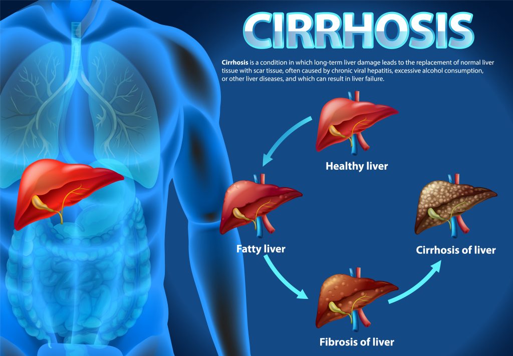
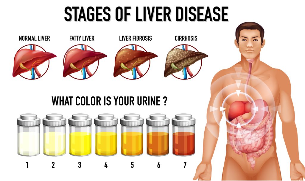
Cirrhosis represents the end-stage of a multitude of chronic liver diseases, characterized histologically by the replacement of normal liver parenchyma with extensive fibrosis and the formation of regenerative nodules. This process is not merely an accumulation of scar tissue but a profound and often irreversible distortion of the liver’s microarchitecture, which fundamentally compromises its vast array of metabolic, synthetic, and detoxification functions. The progression from a healthy liver to a cirrhotic state is a complex, multi-step process driven by chronic necroinflammation, cellular activation, and a dysregulated wound-healing response. Understanding this pathogenic cascade is fundamental to appreciating the clinical manifestations and therapeutic targets of the disease.
From Liver Injury to Fibrosis: The Cellular and Molecular Cascade
The journey to cirrhosis begins with persistent or recurrent injury to the liver. While the nature of the initial insult varies widely—from viral infection and toxic exposure to metabolic overload and autoimmune attack—the liver’s response follows a remarkably conserved pathway of inflammation and fibrogenesis.
The Initial Insult: Hepatocyte Injury and Inflammation
The primary functional cells of the liver, the hepatocytes, are the initial targets in most chronic liver diseases. Chronic exposure to injurious agents leads to hepatocyte stress and death (necrosis or apoptosis). Damaged and dying hepatocytes release a host of intracellular molecules, including reactive oxygen species (ROS) and damage-associated molecular patterns (DAMPs), which function as potent alarm signals within the liver microenvironment. These signals trigger a robust inflammatory response, recruiting immune cells such as neutrophils, monocytes, and lymphocytes to the site of injury. This sustained state of inflammation, or necroinflammation, is the critical engine that drives the subsequent fibrotic process. The continuous cycle of cell death, inflammation, and attempted regeneration creates a pro-fibrogenic milieu that perpetuates liver damage.
The Central Role of Hepatic Stellate Cell (HSC) Activation
The pivotal event in the development of liver fibrosis is the activation of the hepatic stellate cell (HSC). In the healthy liver, HSCs reside in a quiescent state within the perisinusoidal space (the space of Disse), where their primary functions include storing vitamin A and regulating sinusoidal blood flow. Following chronic liver injury, these quiescent cells undergo a dramatic phenotypic transformation into proliferative, contractile, and fibrogenic myofibroblast-like cells.
The Supporting Cast: Kupffer Cells and Sinusoidal Endothelial Cells (SECs)
While HSCs are the primary architects of fibrosis, other non-parenchymal cells play crucial supporting roles. Kupffer cells, the liver’s resident macrophages, are key orchestrators of the inflammatory response. They are activated by DAMPs from injured hepatocytes and, critically, by pathogen-associated molecular patterns (PAMPs), such as lipopolysaccharide (LPS or endotoxin), that translocate from the gut into the portal circulation—a phenomenon exacerbated in conditions like alcohol-related liver disease. Activated Kupffer cells release a cascade of cytokines and chemokines that not only recruit other immune cells but also directly promote the activation and proliferation of HSCs.
Liver sinusoidal endothelial cells (SECs) form the fenestrated lining of the liver sinusoids, facilitating the free exchange of solutes between the blood and hepatocytes. In the setting of chronic injury, SECs lose their characteristic fenestrae, a process known as “capillarization”. This structural change impairs metabolic exchange and contributes significantly to an increase in intrahepatic vascular resistance, laying the groundwork for the development of portal hypertension.
Architectural Distortion: The Transition from Fibrosis to Cirrhosis
Cirrhosis is distinguished from earlier stages of liver disease by a fundamental disruption of the organ’s normal architecture. This is the pathological endpoint where fibrosis progresses from simple scarring to a complete remodeling of the liver lobules.
The Defining Lesion: Regenerative Nodules and Bridging Fibrosis
As fibrogenesis continues unabated, the deposited scar tissue does not remain localized. Instead, it forms fibrous septa that connect, or “bridge,” adjacent portal tracts with each other and with central veins. This bridging fibrosis dissects and partitions the liver parenchyma, entrapping clusters of hepatocytes that are attempting to regenerate. These entrapped, regenerating hepatocyte clusters form the characteristic regenerative nodules that define the cirrhotic liver. The combination of bridging fibrosis and regenerative nodules completely effaces the normal, organized lobular architecture, replacing it with a disorganized, nodular structure that severely impedes both blood flow and hepatocyte function.
Histopathological Classification: Micronodular vs. Macronodular Cirrhosis
The gross and microscopic appearance of the cirrhotic liver can be classified based on the size of the regenerative nodules.
- Micronodular Cirrhosis: Characterized by uniformly small nodules, typically less than 3 mm in diameter, separated by thick, regular fibrous bands. This pattern is often seen in conditions with a persistent, diffuse injurious process, such as alcohol-related liver disease and hemochromatosis.
- Macronodular Cirrhosis: Features nodules of varying sizes, often larger than 3 mm, surrounded by fibrous bands of irregular thickness. This pattern can arise from the coalescence of smaller nodules over time or from insults that cause more extensive, confluent necrosis, such as chronic viral hepatitis.
In many cases, a mixed pattern is observed, and micronodular cirrhosis can evolve into a macronodular form over time.
The Concept of Fibrosis Regression
For many years, cirrhosis was considered an irreversible, end-stage condition. However, a paradigm shift has occurred with the growing recognition that liver fibrosis is a dynamic and potentially reversible process, at least in its earlier stages. If the underlying cause of liver injury is effectively removed or treated—for example, through viral clearance in hepatitis C, sustained alcohol abstinence, or iron depletion in hemochromatosis—the fibrogenic drive ceases. This can lead to a gradual remodeling and resorption of the scar tissue, a process known as fibrosis regression. While the profound architectural distortion of advanced cirrhosis may be permanent, significant improvement in the degree of fibrosis is possible, offering a critical therapeutic window and underscoring the paramount importance of treating the root cause of the disease.
The development of cirrhosis is not a simple linear process of scarring. It involves a self-perpetuating cycle where the consequences of fibrosis fuel further injury. The initial deposition of collagen by activated HSCs, coupled with the capillarization of SECs, creates a structural barrier that increases resistance to blood flow through the liver sinusoids. This is compounded by a functional component, as activated HSCs are contractile and the local production of vasodilators like nitric oxide (NO) is diminished, further constricting the sinusoids. This combined increase in intrahepatic vascular resistance is the primary event that initiates portal hypertension. The resulting high pressure in the portal venous system leads to congestion in the splanchnic circulation and impairs the integrity of the intestinal barrier. This “leaky gut” allows for increased translocation of bacterial products, such as endotoxin, from the intestinal lumen into the portal blood. These endotoxins are potent activators of Kupffer cells and HSCs via Toll-like receptor 4 (TLR4) signaling, which in turn perpetuates the inflammatory and fibrogenic response that caused the portal hypertension in the first place. This establishes a vicious cycle, explaining why cirrhosis, once established, often progresses relentlessly even if the initial insult is controlled. This reframes cirrhosis not merely as a scarring disease but as a complex, integrated disorder of structural fibrosis, progressive vascular dysfunction, and systemic inflammation.
Etiology of Cirrhosis: A Spectrum of Chronic Liver Insults
While the final histopathological outcome of cirrhosis—bridging fibrosis and regenerative nodules—is uniform, the initiating causes are remarkably diverse. A wide range of diseases and conditions can inflict the chronic injury necessary to drive the liver toward this end-stage pathology. Identifying the specific etiology is of paramount clinical importance, as it dictates the primary, disease-modifying therapeutic strategy.
Alcohol-Related Liver Disease (ALD)
Chronic and excessive alcohol consumption is one of the leading causes of cirrhosis worldwide, particularly in Western nations. The pathogenesis of ALD is complex and involves several synergistic mechanisms of injury. The metabolism of ethanol in hepatocytes generates toxic byproducts, most notably acetaldehyde, which can form protein adducts and promote oxidative stress, leading to direct cellular damage. Chronic alcohol use also induces the microsomal enzyme oxidation system (specifically CYP2E1), which generates additional ROS and further contributes to oxidative injury.
A hallmark of ALD is the development of hepatic steatosis, or fatty liver, as alcohol metabolism alters lipid metabolism, promoting fat synthesis and inhibiting its breakdown. Critically, alcohol disrupts the integrity of the intestinal barrier, increasing its permeability. This allows for the translocation of bacterial endotoxins from the gut into the portal circulation, where they trigger a potent inflammatory response by activating Kupffer cells.
The clinical progression typically follows a spectrum from simple steatosis to alcoholic steatohepatitis (ASH), which is characterized by inflammation, hepatocyte ballooning, and the potential presence of Mallory-Denk bodies. It is this inflammatory stage that significantly accelerates the progression to fibrosis and ultimately cirrhosis. Susceptibility varies among individuals, with women generally developing liver damage at lower levels of alcohol consumption than men. Only a fraction of individuals with heavy alcohol use, estimated at 10-20%, will progress to cirrhosis, indicating a significant role for co-factors such as diet, obesity, and genetic predisposition, including variants in the PNPLA3 gene.
Chronic Viral Hepatitis (Hepatitis B, C, and D)
Chronic infection with hepatotropic viruses, particularly hepatitis B virus (HBV) and hepatitis C virus (HCV), is a major global cause of cirrhosis. Unlike the direct toxicity of alcohol, the liver damage in chronic viral hepatitis is predominantly an immune-mediated process. The host’s immune system, in its attempt to clear the virus, mounts a continuous attack on infected hepatocytes. This response, led by cytotoxic CD8+ T cells and Natural Killer (NK) cells, results in a state of chronic low-grade necroinflammation. The persistent cycle of hepatocyte destruction and subsequent regeneration provides a powerful and sustained stimulus for HSC activation and fibrogenesis.
The natural history of these infections is typically indolent, with the progression from chronic infection to clinically significant fibrosis and cirrhosis often taking several decades (10-30 years). However, this timeline can be dramatically accelerated by various co-factors. Concurrent alcohol consumption, co-infection with other viruses (such as HIV or hepatitis D virus in the case of HBV), older age at the time of infection, and metabolic factors like obesity and diabetes all hasten the progression of fibrosis. A key distinction exists in the oncogenic potential of these viruses in the context of cirrhosis: for HCV, hepatocellular carcinoma (HCC) almost invariably develops in a cirrhotic liver, whereas HBV can be directly oncogenic and may cause HCC even in the absence of cirrhosis.
Metabolic Dysfunction-Associated Steatotic Liver Disease (MASLD) and Steatohepatitis (MASH)
Driven by the global epidemics of obesity, type 2 diabetes, and metabolic syndrome, MASLD (formerly non-alcoholic fatty liver disease or NAFLD) has emerged as one of the most common causes of chronic liver disease and a rapidly growing indication for liver transplantation. The disease spectrum begins with metabolic dysfunction-associated steatotic liver (MASL), or simple steatosis, which is the accumulation of fat (triglycerides) in hepatocytes.
In a subset of individuals, this progresses to metabolic dysfunction-associated steatohepatitis (MASH), a more aggressive form of the disease characterized by the addition of inflammation and hepatocyte injury (ballooning degeneration). The transition from MASL to MASH is thought to involve a “second hit,” where factors such as insulin resistance, oxidative stress, cytokine production from adipose tissue, and gut dysbiosis are superimposed on the steatotic liver, triggering necroinflammation. It is the presence of MASH that confers a significant risk for the development of progressive fibrosis, cirrhosis, and HCC. While the progression is generally slow, the sheer prevalence of MASLD makes it a major public health concern. As with other liver diseases, genetic polymorphisms (e.g., in PNPLA3 and TM6SF2) and the severity of underlying metabolic comorbidities are key determinants of disease progression.
Autoimmune and Cholestatic Liver Diseases
This category includes a group of diseases where the primary liver injury is mediated by a dysregulated immune response or is centered on the biliary system.
- Autoimmune Hepatitis (AIH): In AIH, the body’s own immune system loses tolerance to hepatic antigens and launches an attack against hepatocytes. This results in a chronic, fluctuating hepatitis that, if left untreated with immunosuppressive therapy, can rapidly progress to cirrhosis and liver failure. AIH predominantly affects women and is often associated with other autoimmune disorders, such as celiac disease or rheumatoid arthritis.
- Primary Biliary Cholangitis (PBC) and Primary Sclerosing Cholangitis (PSC): These are chronic cholestatic diseases where the primary target of injury is the bile ducts. In PBC, an autoimmune process leads to the progressive destruction of small intrahepatic bile ducts. In PSC, inflammation and fibrosing strictures affect both the intra- and extrahepatic bile ducts, and it is strongly associated with inflammatory bowel disease. In both conditions, the obstruction and impairment of bile flow (cholestasis) lead to the retention of toxic bile acids within the liver. These retained bile acids cause secondary hepatocyte injury, inflammation, and progressive fibrosis, ultimately culminating in a “biliary cirrhosis”.
Genetic and Inherited Disorders
Several inherited metabolic disorders can lead to the accumulation of toxic substances within the liver, causing chronic damage and cirrhosis.
- Hereditary Hemochromatosis: This is an autosomal recessive disorder, most commonly due to mutations in the HFE gene, that results in inappropriately high intestinal iron absorption. The excess iron is deposited in various organs, most notably the liver, where it catalyzes the formation of ROS, leading to oxidative stress, hepatocyte death, and fibrogenesis.
- Wilson’s Disease: A rare autosomal recessive disorder caused by mutations in the ATP7B gene, which is responsible for incorporating copper into ceruloplasmin and excreting excess copper into bile. The genetic defect leads to the toxic accumulation of copper in the liver, brain, and corneas, causing oxidative injury and chronic liver disease that can present as acute liver failure or progress to cirrhosis.
- Alpha-1 Antitrypsin (A1AT) Deficiency: This is an autosomal codominant disorder resulting from mutations in the SERPINA1 gene. The most common severe mutation (Z allele) leads to the production of a misfolded A1AT protein that cannot be efficiently secreted from hepatocytes. This abnormal protein polymerizes and accumulates within the endoplasmic reticulum of hepatocytes, triggering a cellular stress response and a “toxic gain-of-function” injury that results in chronic inflammation, fibrosis, and cirrhosis.
Clinical Presentation and Staging: The Prognostic Watershed
The natural history of cirrhosis is characterized by a long and often clinically silent phase, followed by a transition to a symptomatic phase marked by life-threatening complications. This transition represents the most significant event in the course of the disease, fundamentally altering prognosis and management. The clinical staging of cirrhosis is therefore divided into two distinct phases: compensated and decompensated.
Compensated Cirrhosis: The Asymptomatic Phase
Clinical Presentation
In the compensated stage, the liver, despite extensive scarring and architectural distortion, maintains sufficient functional capacity to prevent the development of overt clinical symptoms. Consequently, patients with compensated cirrhosis are frequently asymptomatic and unaware of their underlying liver disease. The diagnosis is often made incidentally during routine medical evaluations, through the discovery of abnormal liver-related blood tests, or via imaging studies performed for unrelated reasons. When symptoms are present, they are typically mild and non-specific, such as generalized fatigue, malaise, anorexia, or unexplained weight loss, which can easily be attributed to other causes. Physical examination may be entirely normal or may reveal subtle signs such as splenomegaly (an enlarged spleen) or spider angiomata (small, spider-like blood vessels on the skin).
Pathophysiology and Prognosis
Pathophysiologically, compensated cirrhosis is a state where the hepatic functional reserve is preserved. While portal hypertension is typically present, the portal pressure gradient has not yet reached the critical threshold required to cause major clinical sequelae. The prognosis for patients in this stage is relatively favorable, with a median survival time that can exceed 12 years. The primary management goals during this phase are to treat the underlying etiology to prevent further liver damage, to monitor for disease progression, and to conduct surveillance for the development of two key complications: gastroesophageal varices and hepatocellular carcinoma. The presence of varices, even in an asymptomatic patient, is a key prognostic marker that indicates more advanced portal hypertension and a higher likelihood of future decompensation.
Decompensated Cirrhosis: The Onset of Overt Disease
The transition from compensated to decompensated cirrhosis is a pivotal and ominous turning point in the disease’s trajectory. This stage is defined by the development of one or more clinically apparent, major complications of advanced liver disease. The rate of transition from the compensated to the decompensated stage is approximately 5-7% per year.
Defining Decompensating Events
The four cardinal events that define hepatic decompensation are:
- Ascites: The pathologic accumulation of fluid within the peritoneal cavity, leading to abdominal distension and discomfort. It is often the first and most common sign of decompensation.
- Variceal Hemorrhage: Life-threatening bleeding from ruptured gastroesophageal varices, a direct consequence of severe portal hypertension.
- Hepatic Encephalopathy (HE): A spectrum of potentially reversible neuropsychiatric disturbances, ranging from subtle cognitive impairment to confusion, disorientation, and coma, caused by the accumulation of neurotoxins in the brain.
- Jaundice: A yellow discoloration of the skin and sclera due to the liver’s inability to adequately process and excrete bilirubin, reflecting a profound decline in hepatocellular function.
Clinical Presentation and Prognostic Implications
Patients with decompensated cirrhosis are overtly symptomatic and often appear chronically ill. In addition to the defining complications, they may exhibit profound muscle wasting (sarcopenia), malnutrition, easy bruising (coagulopathy), and more pronounced physical signs of portal hypertension, such as caput medusae (distended periumbilical veins).
The development of the first decompensating event is described as a “prognostic watershed” because it signals a dramatic shift in the patient’s prognosis. The median survival plummets from over a decade in the compensated stage to approximately two years after the first decompensating event. The onset of decompensation is a clear indication that the liver can no longer cope with the physiological demands placed upon it and serves as a primary trigger for referral for liver transplantation evaluation.
Diagnostic Evaluation
The diagnosis of cirrhosis requires a comprehensive evaluation that integrates clinical assessment, laboratory data, and imaging findings. The goals of this evaluation are not only to confirm the presence of cirrhosis but also to establish its underlying etiology, assess the degree of functional impairment (staging), and screen for major complications. While liver biopsy remains the definitive gold standard, the diagnostic paradigm has increasingly shifted toward non-invasive methods, which are often sufficient to establish the diagnosis, particularly in the decompensated stage.
Clinical Assessment: History and Physical Examination
A meticulous clinical assessment is the foundational first step in evaluating a patient for suspected liver disease.
- History: The patient’s history can provide crucial clues to the underlying cause of cirrhosis. This includes a detailed quantification of alcohol consumption, a history of risk factors for viral hepatitis (e.g., intravenous drug use, blood transfusions before 1992, high-risk sexual contact), the presence of metabolic risk factors (obesity, diabetes, dyslipidemia), and a family history of liver diseases that might suggest a genetic cause. In the compensated stage, the patient may report only vague symptoms like fatigue, but in the decompensated stage, the history will often include specific complaints such as increasing abdominal girth (ascites), yellowing of the skin (jaundice), mental confusion (hepatic encephalopathy), or vomiting blood (variceal hemorrhage).
- Physical Examination: Physical findings can range from completely normal in well-compensated cirrhosis to strikingly abnormal in advanced disease. Key signs suggestive of chronic liver disease and portal hypertension include splenomegaly (a palpable spleen), palmar erythema (redness of the thenar and hypothenar eminences), and spider angiomata (central arterioles with radiating vessels, typically on the upper torso). As the disease progresses to decompensation, more definitive signs appear, such as jaundice, ascites (detected by shifting dullness or a fluid wave on percussion), peripheral edema, pronounced muscle wasting (especially temporal wasting), and caput medusae (dilated, radiating periumbilical veins).
Laboratory Analysis
Blood tests are essential for assessing the liver’s synthetic function, the extent of hepatocellular injury, and for identifying hematologic abnormalities associated with cirrhosis.
- Liver Function and Synthetic Capacity:
- Aminotransferases (AST and ALT): These enzymes are markers of hepatocellular injury. In cirrhosis, they are often moderately elevated, though they can be within the normal range in inactive, end-stage disease as the number of viable hepatocytes dwindles. A ratio of AST to ALT greater than 2 is highly suggestive of alcohol-related liver disease.
- Bilirubin: Total bilirubin levels are a key indicator of the liver’s excretory function. While typically normal in compensated cirrhosis, bilirubin levels rise as the disease decompensates, leading to clinical jaundice.
- Albumin: As albumin is synthesized exclusively by the liver, its serum concentration is a reliable marker of hepatic synthetic function. Hypoalbuminemia is a hallmark of advanced cirrhosis and contributes to the development of ascites and edema.
- Hematologic and Coagulation Profiles:
- Thrombocytopenia (Low Platelet Count): A platelet count below 150,000/mm³ is one of the most common and earliest laboratory abnormalities in cirrhosis. It is primarily caused by portal hypertension leading to splenomegaly and subsequent sequestration of platelets in the enlarged spleen (hypersplenism), as well as decreased hepatic production of the platelet-stimulating hormone thrombopoietin.
- Prothrombin Time (PT) / International Normalized Ratio (INR): The liver synthesizes the majority of the body’s clotting factors. A decline in synthetic function leads to a prolonged PT and an elevated INR, reflecting impaired coagulation. The INR is a critical, objective measure of liver function and is a core component of the MELD score used for transplant allocation.
- Non-invasive Fibrosis Scores: Several scoring systems have been developed that use routine laboratory values to predict the probability of advanced fibrosis or cirrhosis. The Fibrosis-4 (FIB-4) index (using age, AST, ALT, and platelet count) and the AST to Platelet Ratio Index (APRI) are two commonly used examples. These scores are most valuable for their high negative predictive value, meaning a low score can confidently rule out advanced fibrosis, thereby helping to avoid unnecessary invasive procedures.
Imaging Modalities
Imaging plays a central role in the non-invasive diagnosis of cirrhosis, primarily by assessing the morphological changes of the liver and detecting signs of portal hypertension.
- Abdominal Ultrasound: This is often the initial imaging modality due to its wide availability, non-invasiveness, and low cost. Findings suggestive of cirrhosis include a nodular or irregular liver surface, a coarse or heterogeneous parenchymal echotexture, and changes in liver morphology, such as atrophy of the right lobe and compensatory hypertrophy of the caudate and left lobes. Ultrasound can also readily detect splenomegaly and ascites. Doppler imaging is crucial for assessing the portal vein, including its diameter, flow direction (which can reverse in severe portal hypertension), and patency (to rule out thrombosis).
- Computed Tomography (CT) and Magnetic Resonance Imaging (MRI): These cross-sectional imaging techniques provide superior anatomical detail compared to ultrasound. They can more clearly delineate the nodular liver contour, morphological changes, and the presence of portosystemic collateral vessels (varices). CT and MRI are the primary modalities for the surveillance and characterization of hepatocellular carcinoma, a major complication of cirrhosis.
- Elastography (Transient and MR): These specialized, non-invasive techniques represent a major advancement in the diagnosis and staging of liver fibrosis. Both transient elastography (e.g., FibroScan®) and magnetic resonance elastography (MRE) work by measuring the stiffness of the liver tissue; fibrotic and cirrhotic tissue is significantly stiffer than healthy liver tissue. A high liver stiffness measurement is strongly indicative of advanced fibrosis or cirrhosis. Elastography has demonstrated high accuracy, particularly in excluding or confirming the presence of cirrhosis, and has reduced the need for diagnostic liver biopsy in many patients.
The Role of Liver Biopsy
Procedure
A liver biopsy involves obtaining a small core of liver tissue for histopathological examination. The most common method is a percutaneous biopsy, where a specialized needle is passed through the skin and into the liver, often under ultrasound or CT guidance. Alternative approaches, such as the transjugular route (via the jugular vein) or laparoscopic biopsy, may be used in specific circumstances, such as in patients with severe coagulopathy or ascites. The tissue sample is then processed and examined under a microscope by a pathologist.
Purpose
Historically considered the absolute gold standard for diagnosis, the role of liver biopsy has evolved with the advent of accurate non-invasive tests. Today, it is no longer required for diagnosis in many cases, especially in patients with decompensated disease where clinical, laboratory, and imaging findings are unequivocal. However, biopsy remains an invaluable tool in specific clinical scenarios:
- Diagnostic Uncertainty: When non-invasive tests yield conflicting or indeterminate results.
- Determining Etiology: To diagnose conditions that require histological confirmation, such as autoimmune hepatitis, or to differentiate between MASH and simple steatosis.
- Assessing Disease Activity and Severity: To grade the degree of inflammation and accurately stage the degree of fibrosis, which can influence treatment decisions and prognosis.
- Investigating Other Coexisting Liver Diseases: When more than one cause of liver injury is suspected.
The definitive histological confirmation of cirrhosis on a biopsy is the presence of bridging fibrous septa that divide the parenchyma into regenerative nodules, representing the complete disruption of the normal hepatic architecture.
Systemic Complications and Long-Term Risks
Cirrhosis is a systemic disease that extends far beyond the liver itself. The profound loss of hepatic function and the circulatory derangements of portal hypertension lead to dysfunction in nearly every organ system and predispose patients to a number of severe, long-term risks.
Hepatocellular Carcinoma (HCC) in the Cirrhotic Liver
Pathogenesis and Risk
Cirrhosis is the most potent predisposing risk factor for the development of primary liver cancer, or hepatocellular carcinoma (HCC). A large majority, estimated at up to 90%, of all HCC cases arise in the setting of an underlying cirrhotic liver. The cirrhotic microenvironment is intensely pro-carcinogenic. The relentless cycle of chronic inflammation, hepatocyte death, and compensatory proliferation creates a state of high cellular turnover and oxidative stress, which increases the rate of genetic and epigenetic mutations. The altered signaling pathways, growth factors, and cytokines present in the fibrotic liver further promote the survival and growth of malignant clones. The annual incidence of HCC in patients with established cirrhosis is significant, ranging from 1% to 8% depending on the underlying etiology and other co-factors.
Surveillance Guidelines (NCCN, AASLD)
Given the high risk and the fact that HCC is often asymptomatic in its early, potentially curable stages, surveillance is a standard of care for all patients with cirrhosis. Major professional societies, including the National Comprehensive Cancer Network (NCCN) and the American Association for the Study of Liver Diseases (AASLD), recommend lifelong screening. The standard surveillance protocol consists of an abdominal ultrasound examination performed every six months. This may be combined with the measurement of the serum tumor marker alpha-fetoprotein (AFP), although the utility of AFP is debated due to its limited sensitivity and specificity. The primary goal of surveillance is to detect tumors at an early stage when curative therapies such as surgical resection, ablation, or liver transplantation are still viable options, thereby improving survival.
Hepatorenal Syndrome (HRS) and Acute Kidney Injury (AKI)
Pathophysiology
Patients with advanced cirrhosis are highly susceptible to acute kidney injury (AKI). Hepatorenal syndrome (HRS) is a unique and particularly grim form of AKI that is specific to this patient population. HRS is a functional form of renal failure, meaning it occurs in the absence of any intrinsic structural damage to the kidneys. The pathophysiology is a direct consequence of the extreme circulatory dysfunction of end-stage liver disease. Intense vasodilation in the splanchnic circulation leads to a severe reduction in effective arterial blood volume, which triggers a maximal compensatory activation of systemic vasoconstrictor systems (the RAAS and sympathetic nervous system) in an attempt to maintain blood pressure. This leads to profound renal vasoconstriction, severely reducing renal blood flow and glomerular filtration rate, and culminating in kidney failure.
Management
The management of HRS is challenging and focuses on reversing the systemic vasodilation and improving renal perfusion. This typically involves the administration of intravenous albumin to expand plasma volume, combined with systemic vasoconstrictors such as terlipressin or a combination of midodrine and octreotide. While these medical therapies can serve as a bridge, the only definitive treatment for HRS is liver transplantation, which corrects the underlying circulatory derangement.
Coagulopathy and Hematologic Abnormalities
The liver plays a central role in hemostasis, and cirrhosis leads to complex hematologic derangements.
- Coagulopathy: The traditional view of cirrhotic coagulopathy focused solely on an increased bleeding risk due to the liver’s failure to synthesize pro-coagulant factors (Factors II, V, VII, IX, X) and fibrinogen, which is reflected in an elevated INR. However, it is now understood that the liver also fails to produce key anti-coagulant proteins, such as Protein C, Protein S, and antithrombin. The result is a “rebalanced” but highly unstable hemostatic system, where patients can be at risk for both bleeding and thrombosis (e.g., portal vein thrombosis). The INR, therefore, is a poor predictor of bleeding risk in these patients.
- Anemia and Cytopenias: Anemia is common and multifactorial, resulting from acute or chronic gastrointestinal blood loss, nutritional deficiencies (e.g., folate), bone marrow suppression (particularly in ALD), and hemolysis (spur cell anemia in severe disease). As previously noted, thrombocytopenia is a near-universal finding due to hypersplenism. Leukopenia can also occur for the same reason, compromising the patient’s ability to fight infections.
Malnutrition, Sarcopenia, and Osteoporosis
Metabolic and nutritional complications are universal in patients with advanced cirrhosis and are major contributors to morbidity and mortality.
- Malnutrition and Sarcopenia: Malnutrition is highly prevalent due to a combination of factors, including poor oral intake (due to anorexia, nausea, or early satiety from ascites), malabsorption of dietary fats (due to reduced bile production), and an altered metabolic state where the body is prone to catabolism. Sarcopenia, the progressive loss of skeletal muscle mass and function, is a direct consequence of this and is a powerful independent predictor of complications and mortality in cirrhosis.
- Bone Disease: Patients with cirrhosis, particularly those with cholestatic liver diseases, are at high risk for metabolic bone disease, including osteoporosis and osteopenia. The pathogenesis is multifactorial and includes vitamin D malabsorption and deficiency, hormonal changes, and poor nutrition. This leads to an increased risk of fractures, which can be a significant source of morbidity.

Integrated Management and Therapeutic Strategies
The management of cirrhosis is a multifaceted endeavor that requires a holistic and integrated approach. It is not a single disease to be cured but a complex, chronic condition to be managed. The goals are to treat the underlying cause to prevent progression, proactively manage the complications of liver failure and portal hypertension, accurately assess prognosis to guide major decisions like transplantation, and provide comprehensive supportive care to maintain quality of life.
Management of the Underlying Etiology
The single most important principle in the management of cirrhosis is to identify and treat the root cause of the liver injury. Eliminating the primary insult is the only strategy that can halt the progression of fibrosis, potentially allow for fibrosis regression, and prevent the transition from compensated to decompensated disease. The specific treatment is entirely dependent on the etiology:
- Alcohol-Related Liver Disease (ALD): The cornerstone of treatment is complete and lifelong abstinence from alcohol. This is the most effective intervention for improving outcomes in ALD and is a prerequisite for liver transplant consideration.
- Chronic Viral Hepatitis: For patients with hepatitis C, treatment with direct-acting antiviral (DAA) agents can achieve a cure in over 95% of cases, which has been shown to halt disease progression and reduce the risk of complications. For hepatitis B, long-term therapy with nucleos(t)ide analogues effectively suppresses viral replication, reduces inflammation, and lowers the risk of decompensation and HCC.
- Metabolic Dysfunction-Associated Steatohepatitis (MASH): Management centers on aggressive lifestyle modification, including diet, exercise, and a target weight loss of 7-10%, which can improve steatosis, inflammation, and even fibrosis. The recent FDA approval of resmetirom, a thyroid hormone receptor-beta agonist, marks the first specific pharmacotherapy for MASH with fibrosis.
- Autoimmune Hepatitis (AIH): Treatment involves immunosuppressive therapy, typically with corticosteroids (e.g., prednisone) with or without azathioprine, to control the autoimmune-mediated inflammation.
- Genetic and Inherited Disorders: Management is directed at the specific metabolic defect. This includes therapeutic phlebotomy to remove excess iron in hemochromatosis and the use of chelating agents (e.g., penicillamine) to remove excess copper in Wilson’s disease.
Lifestyle and Nutritional Interventions
Lifestyle and nutritional support are not adjunctive but are core therapeutic pillars in the management of all patients with cirrhosis, particularly those with decompensated disease.
- Dietary Guidelines:
- Sodium Restriction: For patients with ascites or peripheral edema, dietary sodium intake should be restricted to less than 2,000 mg per day to help manage fluid retention. This often requires avoiding processed foods, canned goods, and added salt during cooking.
- Protein Intake: Contrary to outdated advice, protein restriction should be avoided, even in patients with hepatic encephalopathy. Adequate protein intake, targeted at 1.2 to 1.5 grams per kilogram of ideal body weight per day, is crucial to prevent and treat sarcopenia and malnutrition. Protein intake should be distributed throughout the day, with an emphasis on a late-night snack to reduce the overnight fasting period and prevent muscle catabolism. Plant-based and dairy proteins may be better tolerated in some patients with HE.
- Caloric Intake: Patients with cirrhosis are often in a hypermetabolic state and require adequate energy intake (35-40 kcal/kg/day) to prevent malnutrition.
- General Lifestyle Modifications: Complete abstinence from alcohol is mandatory for all patients with cirrhosis, regardless of the initial cause, as alcohol is a direct hepatotoxin. Regular, moderate physical activity, as tolerated, is encouraged to help maintain muscle mass and overall physical conditioning. All medications, including over-the-counter drugs and herbal supplements, should be reviewed by a hepatologist, as many are metabolized by the liver and can be harmful. Non-steroidal anti-inflammatory drugs (NSAIDs) should be strictly avoided due to their risk of precipitating renal failure and gastrointestinal bleeding.
Pharmacological Management of Complications
In addition to treating the underlying etiology, a significant portion of cirrhosis management involves the pharmacological treatment of its complications, as detailed in Section V. This includes:
- Portal Hypertension and Varices: Non-selective beta-blockers (propranolol, nadolol, carvedilol) are used for primary and secondary prophylaxis of variceal bleeding. Vasoactive drugs (octreotide, terlipressin) are used in the acute management of variceal hemorrhage.
- Ascites: Diuretics, specifically a combination of spironolactone and furosemide, are the mainstay of medical therapy.
- Hepatic Encephalopathy: Non-absorbable disaccharides (lactulose) and non-absorbable antibiotics (rifaximin) are used to reduce systemic ammonia levels.
Liver Transplantation
For patients with end-stage liver disease, where medical management is failing and complications are progressing, liver transplantation is the only definitive, life-saving treatment.
- Indications: The primary indications for transplantation are the complications of decompensated cirrhosis (such as refractory ascites, recurrent variceal hemorrhage, or persistent hepatic encephalopathy), progressive liver failure (indicated by a rising MELD score, typically ≥15), or the development of HCC that falls within accepted criteria (e.g., the Milan criteria: one tumor ≤5 cm or up to three tumors each ≤3 cm).
- Evaluation and Outcomes: Potential candidates undergo a rigorous and comprehensive evaluation to assess their medical suitability for the major surgery and their ability to adhere to the complex post-transplant regimen. This includes detailed cardiac, pulmonary, and renal assessments, as well as a thorough psychosocial evaluation. Successful liver transplantation can dramatically improve both survival and quality of life, effectively curing the end-stage liver disease. However, it is a major undertaking that requires lifelong immunosuppressive medication to prevent organ rejection and vigilant management of potential long-term complications such as infection, renal dysfunction, and metabolic disease. The primary limitation to transplantation remains the critical shortage of donor organs.
Future Directions and Emerging Therapies
While current management of cirrhosis focuses on treating the underlying cause and managing complications, the ultimate goal of hepatology research is to develop therapies that can directly halt or even reverse the fibrotic process itself. This represents a paradigm shift from managing the consequences of cirrhosis to treating the disease at its core pathogenic level. Several promising avenues are currently under active investigation.
Regenerative Medicine and Cell-Based Therapies
Regenerative medicine seeks to harness the body’s innate repair mechanisms or to introduce cells that can promote tissue healing and regeneration.
- Stem Cell Therapy: The use of various types of stem cells, particularly mesenchymal stem cells (MSCs), is being investigated for their potential to treat liver fibrosis. It is believed that MSCs exert their therapeutic effects not by differentiating into new hepatocytes, but primarily through paracrine mechanisms—secreting anti-inflammatory, anti-fibrotic, and pro-regenerative factors that modulate the liver microenvironment and promote endogenous repair.
- Macrophage Therapy: A novel and promising approach involves the use of macrophages, which are key immune cells involved in both the progression and resolution of fibrosis. A recent Phase 2 clinical trial (MATCH Phase 2) tested a cell therapy using autologous (patient-derived) macrophages. The results were encouraging, showing a significant reduction in serious liver-related complications in the treated group compared to standard care, suggesting this approach may help stabilize the disease and delay the need for transplantation.
Modulating the Gut-Liver Axis
There is a growing appreciation for the critical role of the “gut-liver axis” in the pathogenesis of chronic liver disease. The translocation of bacterial products from a dysbiotic gut microbiome is a key driver of hepatic inflammation in several etiologies of cirrhosis, including ALD and MASH. Consequently, therapies aimed at modulating the gut microbiome and improving intestinal barrier function are being explored as indirect ways to treat liver disease. These approaches include the use of specific probiotics and prebiotics, as well as fecal microbiota transplantation (FMT), which aims to restore a healthy gut microbial community and reduce the inflammatory burden on the liver.
Patient-Centric Considerations: Quality of Life and Supportive Care
While medical and surgical interventions are the cornerstones of cirrhosis management, a truly comprehensive approach must also address the profound impact the disease has on a patient’s daily life, well-being, and functional status. Improving health-related quality of life (QoL) is a primary goal of therapy, often valued by patients as much as survival itself. This requires a patient-centric model of care that integrates nutritional support, symptom management, and robust psychosocial support systems.
The Burden of Cirrhosis on Quality of Life (QoL)
Living with cirrhosis, particularly in its decompensated stages, imposes a heavy burden on patients. The physical symptoms can be debilitating and include chronic fatigue, persistent pruritus (itching), muscle cramps, and the discomfort and mobility limitations caused by ascites and edema. The cognitive and psychiatric manifestations of hepatic encephalopathy—ranging from subtle “brain fog” and slowed reaction times in minimal HE to overt confusion, personality changes, and sleep disturbances—can be profoundly distressing for both patients and their families, impacting independence, social relationships, and the ability to work or drive. The constant threat of life-threatening complications like variceal bleeding, coupled with the uncertainties of a chronic illness, often leads to significant anxiety and depression.
Nutritional Support as a Therapeutic Pillar
Malnutrition and sarcopenia are not merely complications of cirrhosis; they are integral components of the disease process and powerful independent predictors of adverse outcomes, including infection, hospitalization, and mortality. Therefore, nutritional intervention should be viewed as a core therapeutic strategy, not an afterthought. As detailed previously, this involves ensuring adequate caloric and protein intake to counteract the catabolic state of the disease.
The complexity of balancing these needs with dietary restrictions (such as low sodium for ascites) and patient-specific issues (like anorexia or early satiety) necessitates the involvement of a registered dietitian with expertise in liver disease. A specialized dietitian can perform a detailed nutritional assessment and create a personalized, practical dietary plan that is tailored to the patient’s disease stage, preferences, and lifestyle, thereby improving adherence and clinical outcomes.
